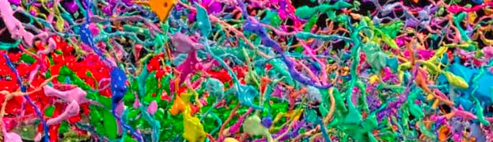
The header image on this post depicts the structure that permits you to read this page, to understand these glyphs as words with a meaning, meaning that is known to you, meaning that is shared with all other humans literate in English, meaning that in some ineluctable way has come to inhabit the brain matter of millions of beings.
It is this convoluted blob of brain structure that permits you to understand the word “permits,” to understand that there exists a referent described by “words,” to recognize exactly what a header image might be, to understand that this Jackson Pollack tangle of colour before you is an exact reproduction of the biological schema that allows you to see the Jackson Pollack tangle of colour before you, to know the meaning of “colour,” “color,” and “Jackson Pollack.”
The header image was obtained from an article on The Guardian Neuroscience page, posted on July 30th, 2015.
The article confirms the resolving power of standard MRI diagnostic imaging as described in a prior post:
Traditional brain imaging techniques, such as MRI, are straightforward to use, but can only resolve features down to about a millimetre. A German anatomical atlas called “BigBrain”, resolves features of the human brain down to micrometres – thinner than a human hair – and almost on a scale of individual cells.
The header image displays brain features measured in millionths of a millimetre. None of what you see in the header image would be revealed in a standard MRI. Standard MRI can only display the most gross characteristics of the brain. MRI typically images areas of blood staining, or areas of the brain devoid of blood due to dead brain cells resulting from a focal injury.
An MRI image only displays the hydrogen content of cellular matter (hydrogen being a component of every water molecule). It does not in fact image the cells themselves. It is a mode of diagnosis by proxy. The doctor who made my diagnosis was operating completely blind. He lacked any valid MRI data upon which to base his medical opinion.
In order to understand my injury you need to know that my brain, something very similar to the coloured tangle you see in the header image above, was impacted by 112,457 ft-lbs of kinetic energy. This energy transfer resulted in a hydrostatic pressure wave within the structure you see in the header. The brain structure was both compressed in upon itself and, due to the high levels of compressive force, oxygen perfusion was inhibited such that elements of the brain micro-structure died, or became unable to perform their normal biologic routine.
I survived the accident. Portions of my brain did not.